


拡散強調MRI (Diffusion-weighted MRI)
侵襲なく、正確ながんの診断と他の画像診断にない機能的診断を可能に
- MRI:磁気の力を利用し、身体内の情報を画像化する検査手法。
- 拡散強調 MRI:組織内の水分子の動きである拡散現象を利用したMRI撮影法。
- 従来のMRI撮影と同時に撮影可能
- 放射線被曝なし
- 造影剤なし、検査前後の安静待機必要なし
- 解剖学的情報に加え、機能的情報を解析可能
がん治療における画像診断には、がんの有無の検索のみならず、質的な評価(がんの悪性度など)、進展度の評価、治療効果の判定が求められ、通常、治療経過中には複数回の画像評価が必要となります。我々は、がんを明瞭に描出する拡散強調MRIを日常診療に応用しています。この検査法は、非侵襲的で、放射線被曝がなく、造影剤を使用しなくても撮影可能な検査です。検査前後の安静待機も必要ありません。拡散強調MRI信号は、がんの有無の診断のみならず、局在や悪性度といった質的な診断に有用であることが示されており、広く臨床使用されるようになっております。
拡散強調MRIは、特に前立腺がんの画像診断において、必須の撮影法となっています。前立腺がんの疑われる方に対する前立腺生検の必要性の評価、生検を行う際に組織採取の標的とするべき部位の同定、前立腺がんの診断がついたのちには、がんの局在を考慮した治療法の選択等に用いられています。
また、腎盂尿管がんや膀胱がんは拡散強調MRIにて明瞭な高信号として描出されます。拡散強調MRIは、時に困難であることもある腎盂尿管がんの診断や、集学的治療を行う筋層浸潤性膀胱がんの各局面における診断に役立てられております。
(拡散強調MRIに興味を持たれた方は下記の「拡散強調MRIの詳細と当科の取り組みについて」をご覧ください。)
拡散強調MRIの詳細と当科の取り組みについて
拡散強調MRIは組織内の水分子の動きである拡散運動を画像化する機能的画像診断法です。1965年にStejskalとTannerにより核磁気共鳴法を使用した拡散計測が報告され、1986年にLe BihanらによりMRIを用いた頭蓋内の拡散現象の画像化が行われました。1990年にMoselyらにより、超急性期の脳梗塞の診断における有用性が示されて以来、頭蓋内病変の診断に臨床利用が進んでおります。
急性期脳梗塞では、細胞浮腫が引き起こされ、細胞外液腔が狭くなります。そのため細胞外液腔の水分子の動きが制限されるため、周囲の脳実質と比較して高信号を示すことになります。同様に、がん組織では細胞密度が高く、細胞間隙が狭くなり、強い高信号を示します。一方、周囲臓器の信号は抑制されるため、がん組織と周囲組織の信号の間には良好なコントラストが生じることになります。そのため、拡散強調MRIはがん組織の診断に優れております。さらに、化学療法や放射線療法により、がん組織の細胞密度の低下や細胞膜の破壊が生じると、水分子の動きの制限が弱くなり、拡散強調MRI信号は減弱もしくは消失することになります。治療による拡散強調MRI信号の変化は治療効果や病勢を反映するため、拡散強調MRIは治療中の経過観察に優れています。
当科では悪性疾患の診断・治療において、正確な診断を非侵襲的に行うべく日々検討をおこなっております。拡散強調MRIの有用性に早い段階より注目し、泌尿器癌の診療における臨床応用を進めてまいりました。その有用性については下記の国際医学雑誌に報告を行なっており、高い評価を頂いております。
腎盂尿管がんの診断における拡散強調MRIの有用性については、当教室が世界に先駆けて報告しており、2010年度欧州泌尿器科学会・腫瘍学部門で 3rd prize を受賞しました。2010年に報告した膀胱がんに対する化学放射線療法の治療効果判定における有用性については、欧州泌尿器科学会の筋層浸潤膀胱がんの治療ガイドラインにも取り上げられております。また、2016年には、定量的に評価した拡散強調MRI信号により、腎盂尿管がんの予後が層別化されることを確認し、米国泌尿器科学会にてBest presentation awardを受賞しております。2017年には、AJR誌に掲載された膀胱がんのImaging biomarkerとしての有用性についての総説は、米国放射線科学会が発行するIn practice誌のSummer 2017号に取り上げられております。
拡散強調MRIと前立腺生検の結果を組み合わせた、前立腺癌の診断法は、国際的に高い評価を受けております。2012年度欧州泌尿器科学会におけるBest presentation awardや本邦の泌尿器科学会の総会賞および泌尿器科学会賞を受賞しております。
-
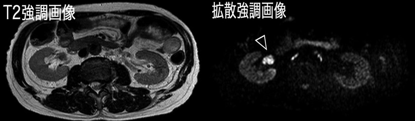
右腎盂がん (Grade 3, pT3) のMRI画像。拡散強調MRI画像において、右腎盂を占める腫瘍は、周囲の抑制された信号を背景に、明瞭な高信号を示している。T2 強調MRI画像と比較して、拡散強調MRI画像では病変をより明確に指摘可能となっている。
実績
Award
欧州泌尿器科学会腫瘍部門3rd prize
Yoshida S, Masuda, H, Ishii, C, Tanaka, H, Komai, Y, Numao, N, Saito, K, Koga, F, Fujii, Y, Kawakami, S, Kihara, K. Diffusion-weighted magnetic resonance imaging in diagnosis of upper urinary tract cancer. "Third Prize for the Best Abstract (Oncology)" EAU Annual Congress, Barcelona, 2010/4/17-
欧州泌尿器科学会ベストポスター賞
Yoshida S, Masuda, H, Ishii, C, Tanaka, H, Komai, Y, Numao, N, Saito, K, Koga, F, Fujii, Y, Kawakami, S, Kihara, K. Diffusion-weighted magnetic resonance imaging in diagnosis of upper urinary tract cancer. "Best Poster Presentation" EAU Annual Congress, Barcelona, 2010/4/17
-
欧州泌尿器科学会ベストポスター賞
Matsuoka Y, Numao N, Saito K, Tanaka H, Kijima T, Kobayashi S, Tatokoro M, Sakura M, Yokoyama M, Ishioka J, Koga F, Masuda H, Kihara K. Candidate selection for hemiablative focal therapy of prostate cancer through a combination of extended 14-core biopsy and MRI. "Best Poster Presentation" The 27th Annual Congress of the European Association of Urology, Paris, France, 2012/2/25.
-
International Symposium on Focal Therapy and Imaging in Prostate & Kidney Cancer: 3rd prize of best poster award
Matsuoka Y, Numao N, Saito K, Tanaka H, Ito M, Yoshida S, Yokoyama M, Ishioka J, Fujii Y, Kihara K. Eligibility analysis for focal therapy based on prostatectomy findings: Does intermediate-risk cancer have a higher likelihood of undertreatment than low-risk cancer?, "3rd prize of best poster award". 8th International Symposium on Focal Therapy and Imaging in Prostate & Kidney Cancer. Amsterdam, 2015/6/21
-
米国泌尿器科学会ベストポスター賞
S. Yoshida, Y. Uchida, S. Kobayashi, F. Koga, H. Tanaka, M. Yokoyama, J. Ishioka, Y. Matsuoka, N. Numao, K. Saito, Y. Fujii, K. Kihara. “#MP27 Apparent diffusion coefficient as a prognostic biomarker of upper urinary tract cancer”, "Best Poster Presentation", 110th annual meeting of the American Urological Association, San Diego, USA, 2016/5/7
-
第100回日本泌尿器科学会総会 総会賞
松岡 陽、沼尾 昇、齋藤一隆、田中 宏、木島敏樹、小林秀一郎、田所 学、砂倉瑞明、横山みなと、石岡淳一郎、古賀文隆、増田 均、木原和徳. 前立腺14ヵ所生検とMRIは片側部分治療の適格症例を高い精度で予測する.
第100回日本泌尿器科学会総会 横浜, 2012/4/21 -
第103回日本泌尿器科学会総会 総会賞
松岡 陽、沼尾 昇、田中 宏、伊藤 将也、中山 貴之、井上 雅晴、田所 学、吉田 宗一郎、横山 みなと、石岡 淳一郎、齋藤 一隆、藤井 靖久、木原 和徳. focal therapyは高リスク前立腺癌へ適用可能か:MRIと生検による適格性診断能のリスク群別解析. 第103回日本泌尿器科学会総会 2015年4月18日
-
第21回日本泌尿器科学会賞
松岡 陽
Combination of diffusion-weighted magnetic resonance imaging and extended prostate biopsy predicts lobes without significant cancer: application in patient selection for hemiablative focal therapy. -
第623回日本泌尿器科学会東京地方会ベストプレゼンテーション賞
矢嶋 習吾、吉田 宗一郎、横山 みなと、石岡 淳一郎、松岡 陽、齋藤 一隆、藤井 靖久. 全身拡散強調MRIが活動性骨転移病巣の同定と治療効果の判定に有用であった去勢抵抗性前立腺癌の2例. 第623回日本泌尿器科学会東京地方会、2017年2月16日
-
第107回 日本泌尿器科学会総会 総会賞
内田 裕將、吉田 宗一郎、下田 海生、田中 宏、阪本 剛、上原 翔、安田 庸輔、木島 敏樹、横山 みなと、石岡 淳一郎、松岡 陽、齋藤 一隆、藤井 靖久. 拡散強調MRIのwhole-lesion ADC 解析を用いた嫌色素性腎癌と腎オンコサイトーマの鑑別. 第107回 日本泌尿器科学会総会 2019年4月19日
Textbook
Yoshida S, Masuda H, Koga F, Tanaka H and Kihara K: Functional CT and MRI of the Urinary System and Adrenal Glands: Functional Imaging in Oncology, Springer, 2014, pp 1173-1182.
Invited Review
Yoshida S, Koga F, Kobayashi S, et al.: Diffusion-weighted magnetic resonance imaging in management of bladder cancer, particularly with multimodal bladder-sparing strategy. World J Radiol. 6: 344-54, 2014.
Yoshida S, Koga F, Masuda H, Fujii Y and Kihara K: Role of diffusion-weighted magnetic resonance imaging as an imaging biomarker of urothelial carcinoma. Int J Urol. 21: 1190-200, 2014.
Yoshida S, Takahara T, Kwee TC, Waseda Y, Kobayashi S and Fujii Y: DWI as an Imaging Biomarker for Bladder Cancer. AJR Am J Roentgenol: 1-11, 2017.
Publication
前立腺
Numao N, Yoshida S, Komai Y, et al.: Usefulness of pre-biopsy multiparametric magnetic resonance imaging and clinical variables to reduce initial prostate biopsy in men with suspected clinically localized prostate cancer. J Urol. 190: 502-8, 2013.
Bae H, Yoshida S, Matsuoka Y, et al.: Apparent diffusion coefficient value as a biomarker reflecting morphological and biological features of prostate cancer. Int Urol Nephrol. 46: 555-61, 2014.
Matsuoka Y, Numao N, Saito K, et al.: Combination of diffusion-weighted magnetic resonance imaging and extended prostate biopsy predicts lobes without significant cancer: application in patient selection for hemiablative focal therapy. Eur Urol. 65: 186-92, 2014.
Matsuoka Y, Numao N, Saito K, et al.: Candidate selection for quadrant-based focal ablation through a combination of diffusion-weighted magnetic resonance imaging and prostate biopsy. BJU Int. 117: 94-101, 2016.
Matsuoka Y, Ishioka J, Tanaka H, et al.: Impact of the Prostate Imaging Reporting and Data System, Version 2, on MRI Diagnosis for Extracapsular Extension of Prostate Cancer. AJR Am J Roentgenol: W1-W9, 2017.
Waseda Y, Yoshida S, Takahara T, et al.: Utility of computed diffusion-weighted MRI for predicting aggressiveness of prostate cancer. J Magn Reson Imaging. 46: 490-496, 2017.
尿路上皮癌
Yoshida S, Masuda H, Ishii C, Saito K, Kawakami S and Kihara K: Initial experience of functional imaging of upper urinary tract neoplasm by diffusion-weighted magnetic resonance imaging. Int J Urol. 15: 140-3, 2008.
Yoshida S, Koga F, Kawakami S, et al.: Initial experience of diffusion-weighted magnetic resonance imaging to assess therapeutic response to induction chemoradiotherapy against muscle-invasive bladder cancer. Urology. 75: 387-91, 2010.
Kobayashi S, Koga F, Yoshida S, et al.: Diagnostic performance of diffusion-weighted magnetic resonance imaging in bladder cancer: potential utility of apparent diffusion coefficient values as a biomarker to predict clinical aggressiveness. Eur Radiol. 21: 2178-86, 2011.
Yoshida S, Masuda H, Ishii C, et al.: Usefulness of diffusion-weighted MRI in diagnosis of upper urinary tract cancer. AJR Am J Roentgenol. 196: 110-6, 2011.
Yoshida S, Koga F, Kobayashi S, et al.: Role of diffusion-weighted magnetic resonance imaging in predicting sensitivity to chemoradiotherapy in muscle-invasive bladder cancer. Int J Radiat Oncol Biol Phys. 83: e21-7, 2012.
Yoshida S, Kobayashi S, Koga F, et al.: Apparent diffusion coefficient as a prognostic biomarker of upper urinary tract cancer: a preliminary report. Eur Radiol. 23: 2206-14, 2013.
Kobayashi S, Koga F, Kajino K, et al.: Apparent diffusion coefficient value reflects invasive and proliferative potential of bladder cancer. J Magn Reson Imaging. 39: 172-8, 2014.
Uchida Y, Yoshida S, Kobayashi S, et al.: Diffusion-weighted MRI as a potential imaging biomarker reflecting the metastatic potential of upper urinary tract cancer. Br J Radiol. 87: 20130791, 2014.
Nakamura Y, Yoshida S, Tanaka H, et al.: Potential Utility of Diffusion-Weighted Magnetic Resonance Imaging in Diagnosis of Residual Bladder Cancer before Second Transurethral Resection. Urol Int. 98: 298-303, 2017.
Nishizawa T, Yoshida S, Koga F, et al.: Standardization of the apparent diffusion coefficient value of bladder cancer across different centers: Applicability in predicting aggressive pathologic phenotypes. Clin Imaging. 44: 121-126, 2017.
吉田 宗一郎, 増田 均.【腎・泌尿器癌 基礎・臨床研究のアップデート】 膀胱癌 臨床研究 診断と分類 画像診断. 日本臨床. 2010; 68: 245-248.
中山貴之、吉田宗一郎、藤井靖久ら. 膀胱癌リンパ節転移に対するMRI拡散強調画像の使用経験 -化学療法効果判定における可能性について- 日泌尿会誌. 2008; 99: 737-741.
森山真吾, 吉田宗一郎, 竹下英毅ら. MRI拡散強調画像陽性を呈した膀胱炎症性病変の1例;組織学的所見との比較検討による考察. 日泌尿会誌. 2013; 104: 545-548.
中村祐基、吉田宗一郎、森山慎吾ら. TURBTの術後変化と残存浸潤癌との鑑別に拡散強調MRIが有用であった1例.泌尿器外科. 2015; 28: 1113-1116.
Yajima S, Yoshida S, Takahara T, et al. Usefulness of the inchworm sign on DWI for predicting pT1 bladder cancer progression. Eur Radiol. 2019 Mar 19. doi: 10.1007/s00330-019-06119-6.
その他
Yoshida S, Fujii Y, Yokoyama M, et al.: Female urethral diverticular abscess clearly depicted by diffusion-weighted magnetic resonance imaging. Int J Urol. 15: 460-1, 2008.
Tanaka H, Yoshida S, Fujii Y, et al.: Diffusion-weighted magnetic resonance imaging in the differentiation of angiomyolipoma with minimal fat from clear cell renal cell carcinoma. Int J Urol. 18: 727-30, 2011.
Yachida Y, Yoshida S, Takeshita H, et al.: Bone abnormal signal incidentally found in pre-biopsy diffusion-weighted MRI for suspected prostate cancer: what does it reflect? Urol Int. 93: 170-5, 2014.
吉田 宗一郎, 木原 和徳.【腎機能と画像診断】 血尿の腎画像診断の考え方. 成人病と生活習慣病 2008; 38: 803-808.